

File:TransientReceptorPotential-channel.png

Original file (2,799 × 1,094 pixels, file size: 660 KB, MIME type: image/png)
Summary[edit | edit source]
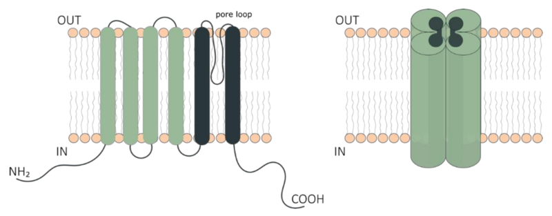
TRP channel
Schematic representation of Transient Receptor Potential (TRP) ion channel structure. The six-transmembrane segment topology of the monomer (left) and tetrameric functional unit of TRP channels (right). The cytoplasmic NH2- and COOH-terminal domains, and the transmembrane domain constituting the pore region are indicated. The transmembrane domain comprises segments 1–4 (green) distal to the pore and the segments 5 and 6 (dark green) proximal to the pore of the channel.
Author: Boonen et al (2018. Citation: Toxins 2018, 10(8), 326; https://doi.org/10.3390/toxins10080326 Source: TRP Channels as Sensors of Bacterial Endotoxins Fig 1 https://www.mdpi.com/2072-6651/10/8/326
Licensing[edit | edit source]
 |
This file is licensed under the Creative Commons Attribution 4.0 International license. | |
|
File history
Click on a date/time to view the file as it appeared at that time.
| Date/Time | Thumbnail | Dimensions | User | Comment | |
|---|---|---|---|---|---|
| current | 12:00, May 11, 2019 | 2,799 × 1,094 (660 KB) | Notjusttired (talk | contribs) | TRP channel Author: Boonen et al (2018. Citation: Toxins 2018, 10(8), 326; https://doi.org/10.3390/toxins10080326 Source: TRP Channels as Sensors of Bacterial Endotoxins Fig 1 https://www.mdpi.com/2072-6651/10/8/326 |
You cannot overwrite this file.
File usage
The following page uses this file: